[技術分享] Cytology
Quantitative phase imaging for cytology
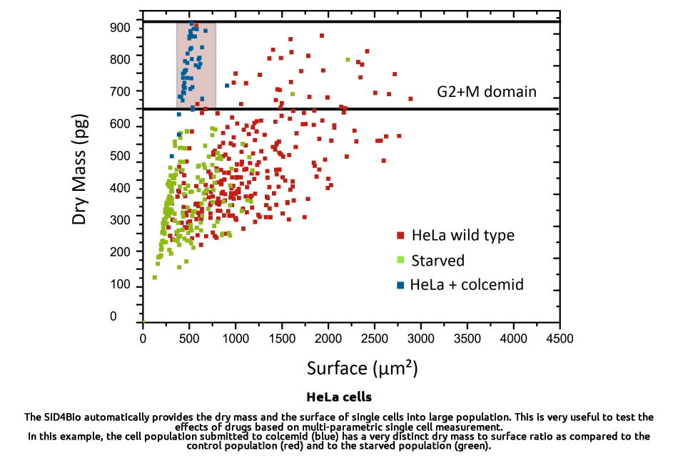
The Phasics quantitative phase microscopy solution allows analyzing large living-cell populations at the single-cell level. It delivers a comprehensive dataset of accurate quantitative parameters for individual cells such as: morphology (surface, shape factor…), dry mass, and many phase-shift-related parameters (density, homogeneity, proteins distribution… ). Thus it is ideal for multiple assays as an automated image cytometer.
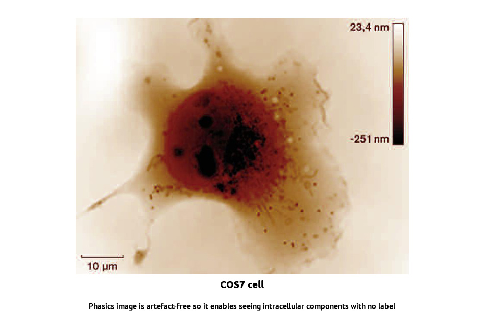
- Artefact-free images: ensure robust and automated segmentation and measurements
- Label-free technique: allows long time-lapse microscopy for non-invasive cellular study: motility, proliferation, monitoring of cell cycle, apoptosis, viability, differentiation, cytotoxicity…
- Merging with fluorescence microscopy: to get a comprehensive dataset at the cellular and molecular level
- Easy integration to high-content screening platform: provides valuable quantitative data combined with machine learning algorithms to enable automated diagnosis
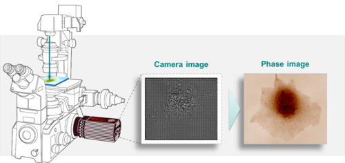
▽ Measurement setup
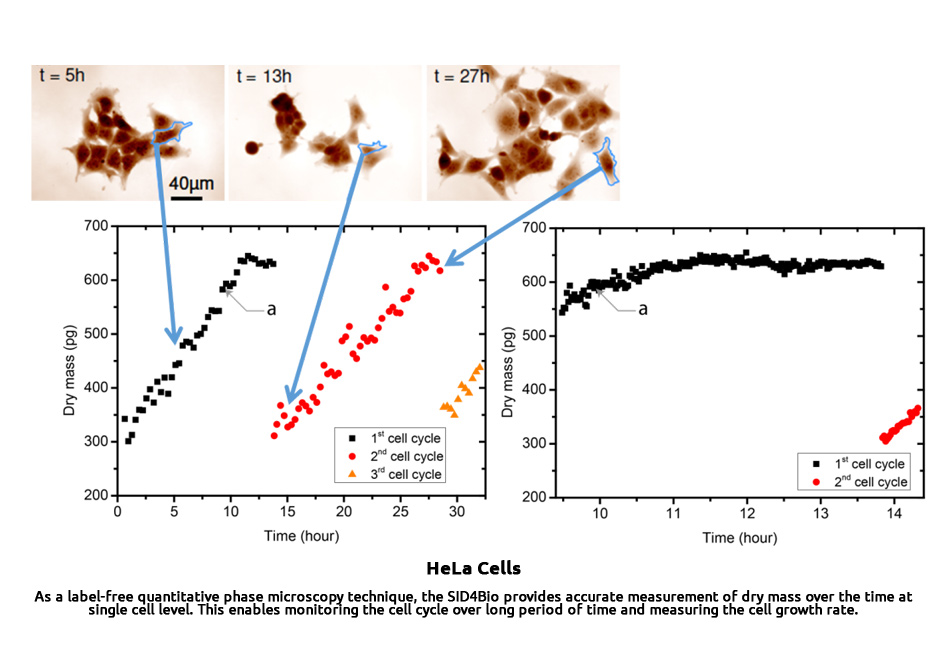
Dry mass and morphometric parameters are proven indicators of many cell mechanisms: viability (apoptosis detection…), cell growth (cell cycle status, proliferation…), and cell differentiation based on phenotypic characteristics. They also reveal cell abnormalities: shape change, heterogeneity, presence of parasites… They are useful parameters to identify and monitor cells in a large population.
It can be applied to:
- Cancer cell proliferation & growth rate monitoring
- Pharmacology research: drug screening, drug discovery, cytotoxicity assays
- Bioprocess: Cell culture monitoring, microbiology
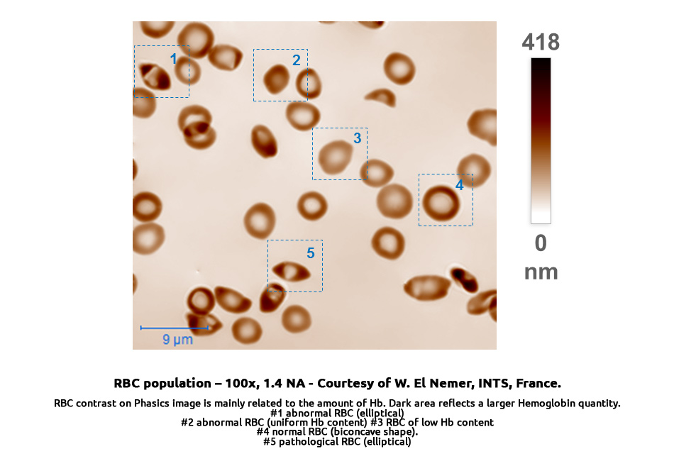
- Blood testing: Red blood cell pathology identification such as anemia type identification, parasitemia calculation
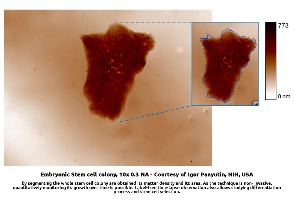
- Stem cell monitoring & selection in regenerative medicine
▽ Applications gallery
Cell growth and apoptose for cancer reseach
Bio production and microbiology
Research
Blood testing
Drug testing and toxicology